In recent years, the exploration of neural circuits has become increasingly sophisticated, with scientists employing genetically encoded voltage indicators (GEVIs) to assess the electrical activity of neurons. These indicators are vital tools that enable researchers to decipher how neurons transmit information and interact within the intricate web of the brain’s architecture. As neuroscientists advance in this area, they face the ongoing debate over the efficacy of one-photon (1P) versus two-photon (2P) voltage imaging techniques. A recent study from Harvard University provides critical insights into the advantages and limitations associated with each method, offering a foundation for future neural imaging initiatives.
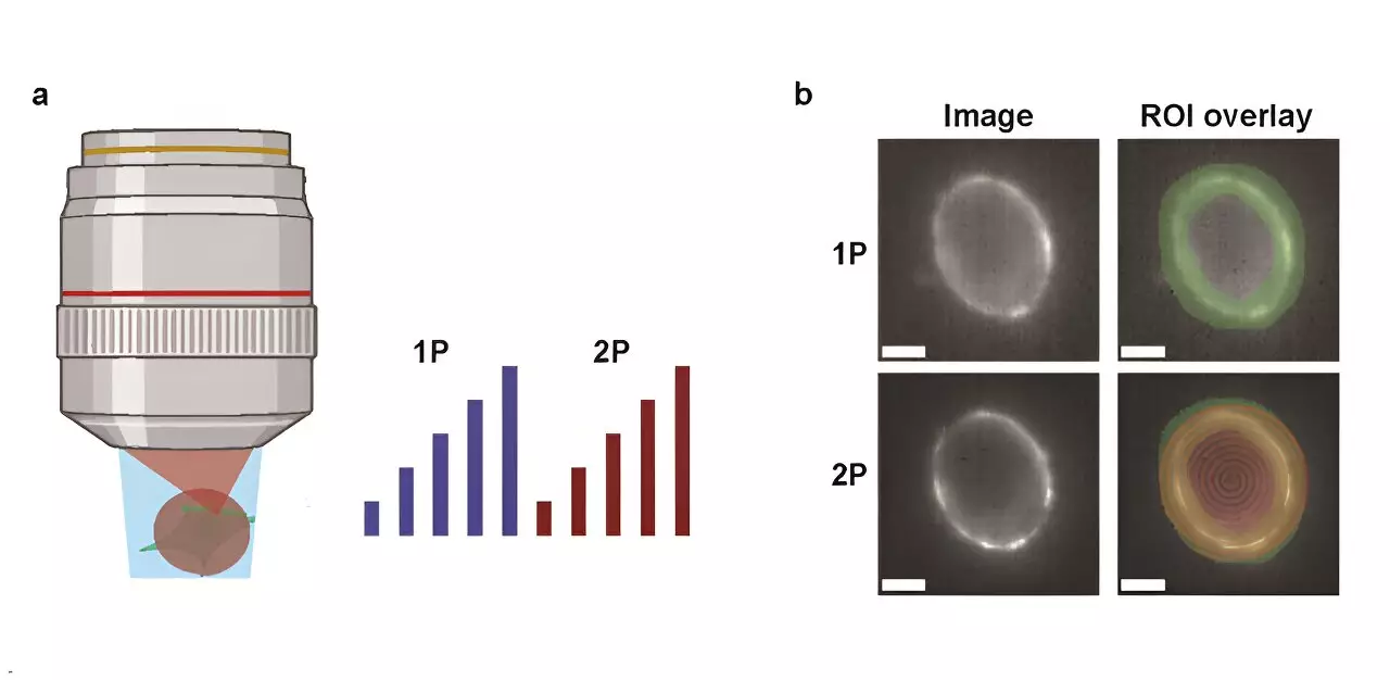
The research, published in Neurophotonics, undertook a detailed comparative analysis of 1P and 2P voltage imaging. The focus was on the optical characteristics and biophysical constraints inherent to each approach. The authors meticulously measured key parameters, including the brightness levels and voltage sensitivity of popular GEVIs under both imaging modalities. This evaluation is crucial because variations in fluorescence with brain depth significantly influence the viability of in vivo imaging techniques.
To quantify their observational findings, the research team developed an innovative model that predicts the possible number of neurons that can be accurately captured based on the properties of the reporters used, the imaging parameters, and required signal-to-noise ratios (SNR). This modeling adds a layer of complexity to the analysis, emphasizing that successful neuronal measurement relies not only on the indicators but also on the methodology employed.
A pivotal discovery highlighted in the study is that the illumination power demand for 2P excitation is drastically higher—approximately 10,000 times more per cell compared to 1P illumination—for achieving similar photon count rates. This requirement opens a can of challenges for researchers: it increases the risk of photodamage to neural tissue and introduces issues related to shot noise. For example, in experiments employing the JEDI-2P indicator in the cortex of mice, researchers found that, given a laser power cap of 200 mW and aiming for a SNR of 10 with a repetition rate of 80MHz, they could only monitor around twelve neurons located deeper than 300 micrometers.
Such limitations raise important questions about the practicality of 2P voltage imaging in live models. The combination of the photon-count requirements and the moderate voltage sensitivity of existing GEVIs makes it challenging to achieve optimal imaging outcomes, particularly when the objective is to collect data from a vast number of neurons at substantial depths.
The study emphasizes the delicate balance between the benefits and trade-offs of employing 1P versus 2P techniques for cellular voltage imaging. It also shines a light on the pressing need for innovation in this field. Continuous enhancements in sensor technology and next-generation imaging methods will be essential to resolve the existing challenges. The findings from this research present a roadmap for how future developments can enhance our comprehension of neural circuits and neuronal communication processes.
As the scientific community pushes forward, leveraging the insights from such studies will prove vital for making tangible advancements in neural circuitry research, potentially paving the way for groundbreaking discoveries in neuroscience.
Leave a Reply