Recent advancements at the University of California, Los Angeles (UCLA) have propelled the field of imaging technology forward in a remarkable way. The introduction of a wavelength-multiplexed diffractive optical processor represents a paradigm shift in 3D Quantitative Phase Imaging (QPI), a technique that has traditionally faced significant challenges. Conventional QPI methods often require complex setups involving diverse illumination angles and extensive digital post-processing, making them cumbersome and labor-intensive. The innovative processor developed by UCLA researchers promises to streamline this process, ushering in a new age of high-resolution imaging that is not only efficient but also practical for real-world applications like biomedical diagnostics and materials science.
Unveiling Complex Structures with Clarity
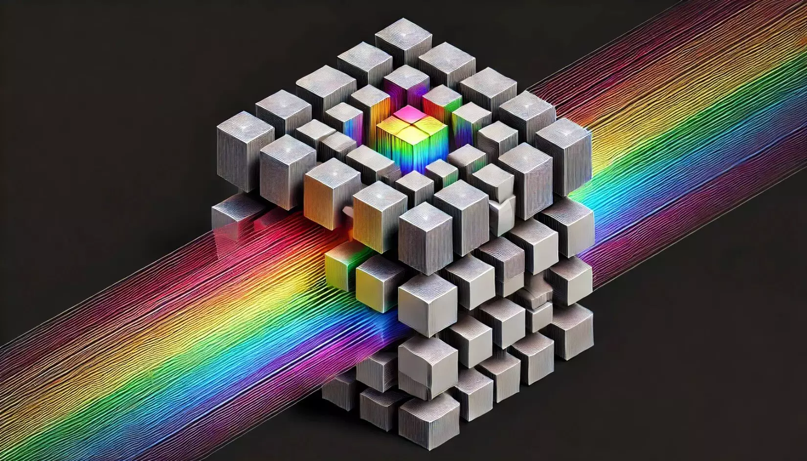
Traditional imaging methods struggle with accurately capturing the minute details of weakly scattering samples. This hampers the ability to visualize transparent specimens, which are prevalent in biological studies. The strength of the new wavelength-multiplexed diffractive optical processor lies in its ability to convert complex phase distributions into distinct intensity patterns encoded at different wavelength channels. This approach allows for simultaneous imaging of multiple objects situated at varying axial positions, radically increasing the information captured in a single operation. This leap will play a crucial role in enhancing the resolution and clarity of images, fostering breakthroughs in diagnosing diseases and analyzing intricate biological structures.
Efficiency Merges with Precision
The innovation showcases the prowess of deep learning, applied to optimize passive diffractive optical elements for phase-to-intensity transformations. The simultaneous imaging of distinct phase objects without needing intricate digital algorithms paves the way for highly efficient QPI solutions that maintain robustness and precision. The compact nature of the optical processor signifies a substantial improvement over bulky setups traditionally required for high-quality phase imaging. Moreover, the capability of performing quantitative phase imaging rapidly across multiple axial planes suggests new pathways for real-time imaging applications—an essential factor for fields that require immediate analysis, such as emergency diagnostics in hospitals and critical environmental assessments.
Broad Applications and Expanding Horizons
Notably, the implications of this research extend beyond mere laboratory application. The design’s adaptability to various regions of the electromagnetic spectrum—including visible light and infrared—opens doors for integration into a wide range of imaging systems. This expansion hints at futuristic applications in environmental monitoring, materials characterization, and far-reaching biomedical applications. The ability to deploy this technology on a more extensive scale and adapt it to specialized needs makes it a potent tool for scientists and researchers across multiple disciplines.
UCLA’s innovative leap in 3D QPI not only addresses the limitations of traditional methods but also sets the stage for future advancements in imaging technology. With the potential for high-resolution, label-free imaging of transparent samples, it holds promise for revolutionary changes in diagnostics and beyond. The world now stands at the beginning of an exciting journey into the depths of visualization across various scientific frontiers, made possible through the integration of advanced optical processing and deep learning technologies.
Leave a Reply